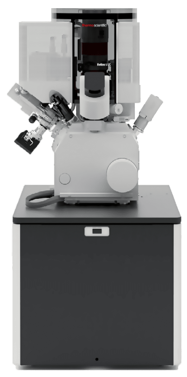
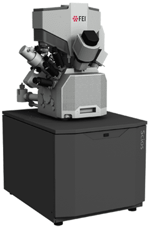
Focused Ion Beam Scanning Electron Microscopy (FIB-SEM)

Like other high-resolution scanning electron microscopes, Focused-ion-beam scanning electron microscopes (FIB-SEMs) are used to produce 2D and 3D images of surface topography and can resolve nm-scale features on a sample surface.

The FIB allows advanced analytical workflows such as cross-section, tomography, lithography, lamella prep, etc.

Strengths

  • Ultra-high resolution imaging capabilities (limit resolution is < 1 nm)
  • Able to image multi-modal, sub-surface, and 3D information
  • FIB enables precise manipulation of sample in-situ: cutting, cleaving, trenching, exposing, and ion-welding of different fragments on the sample for imaging and analysis

Limitations

  • Analysis is destructive
  • Insulating materials impair precision of ion beam action and reduce imaging resolution

Learn More

Focused Ion Beam Scanning Electron Microscopy Services

Focused Ion Beam Scanning Electron Microscopy

Like other high-resolution scanning electron microscopes, Focused-ion-beam scanning electron microscopes (FIB-SEMs) are used to produce 2D and 3D images of surface topography and can resolve nm-scale features on a sample surface.

The FIB allows advanced analytical workflows such as cross-section, tomography, lithography, lamella prep, etc.

Sample Requirements

Example Outputs

FIB-SEM cross-section of a pixel array, showing underlying nm-scale device features.

Instruments Used

ThermoFisher Scientific Helios 5 UC (x3)

ThermoFisher Scientific Helios 5 UC (x3)

  • Maximum Horizontal Field Width: 2.3 mm at 4 mm WD
  • Electron Beam:
    • Resolution Limit: 0.7 nm at 1 kV
    • Current Range: 0.8 pA to 100 nA
    • Accelerating Voltage Range: 350 V to 30 kV
  • Ion Beam:
  • Electron Beam:
    • Resolution Limit: 4.0 nm at 30 kV using preferred statistical method
    • Current Range: 1 pA to 100 nA
    • Accelerating Voltage Range: 500 V to 30kV
View Instrument Brochure
ThermoFisher Scientific Helios 5 UX

ThermoFisher Scientific Helios 5 UX

The features of the Helios 5 UC, di

  • Phoenix Ion Column with superior high-current and low-voltage performance
    • Ion beam current range: 1 pA – 65 nA
    • Accelerating voltage range: 500 V – 30 kV
    • Time-of-flight (TOF) correction
  • The Helios 5 UX DualBeam features an ultra-high-brightness electron source with next-generation UC+ monochromator technology to reduce the beam energy spread below 0.2 eV for beam currents up to 100 pA
View Instrument Brochure
ThermoFisher Scientific Scios

ThermoFisher Scientific Scios

Optimized to achieve best performance across a wide array of sample types.

  • Powerful charge neutralization
  • Enables analysis on magnetic samples
  • Able to operate above vacuum pressure

How FIB-SEM Works

On a FIB-SEM, the added focused-ion-beam allows for in situ sample manipulation. Normally the FIB beam is used to cross-section the sample at a precise location, but it can accomplish many other tasks such as: tomography, lithography, lamella prep, and more.

The imaging capabilities of the scanning-electron-beam in a FIB-SEM work as they do in any other SEM: the system generates an image by detecting electrons scattered by a highly-focused, high-energy applied electron beam as it is raster-scanned over the surface of a sample.

The secondary focused-ion-beam is comprised of high-energy, charged atoms (most commonly Ga+). When applied to the sample, the FIB can be tuned to either ablate material from the surface, or deposit atoms of an accompanying neutral gas.

Additionally, both in-house FIB-SEM instruments at Covalent also incorporate energy dispersive spectroscopy (EDS) detectors that enable measurement and mapping of elemental composition alongside all other FIB-SEM operations.