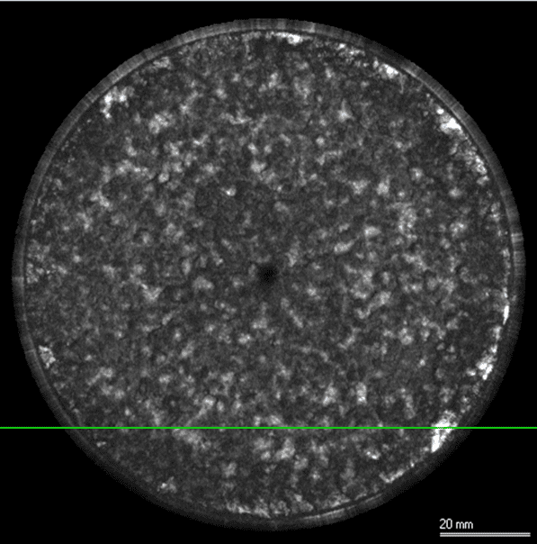
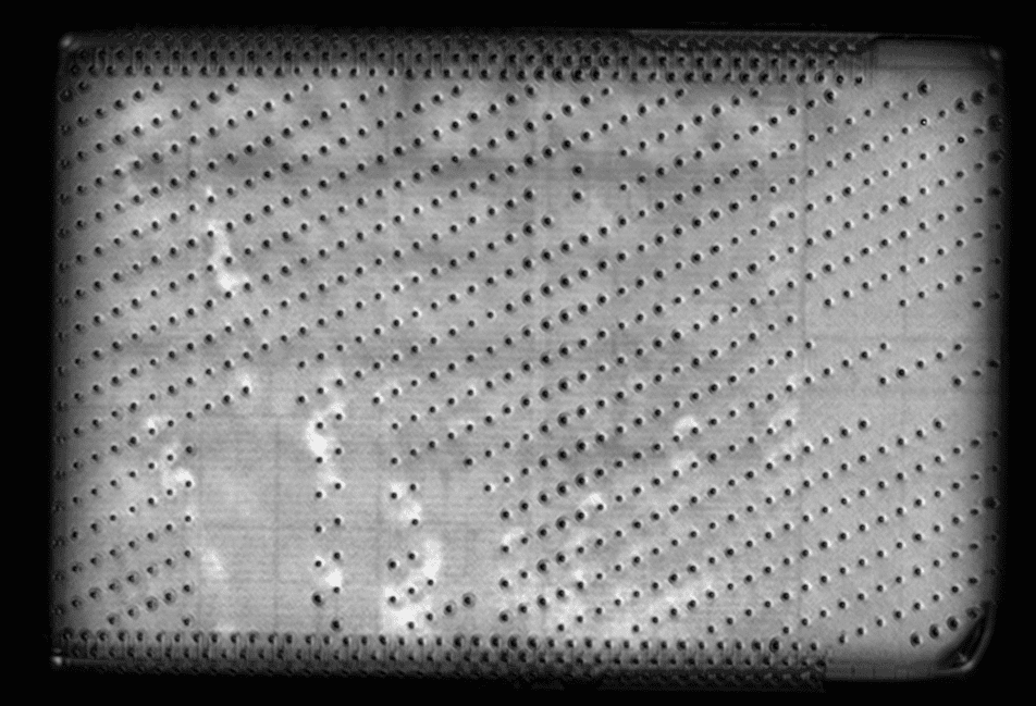
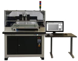
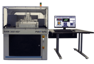
Scanning Acoustic Microscopy (SAM)
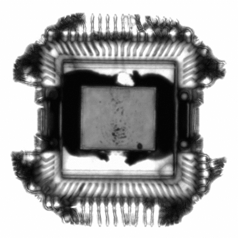
Scanning Acoustic Microscopy (SAM) is a non-destructive and non-invasive imaging technique that uses ultrasound signals to visualize the internal structures of a sample.
Strengths
- High penetration depth enables visualization of underlying and internal structures
- Able to characterize buried topographies which are difficult or impossible to resolve with other microscopy techniques
- Non-destructive analysis, however sample will get wet
Limitations
- Slower processing time than micro-CT
- Reduced spatial resolution compared to electron microscopy techniques