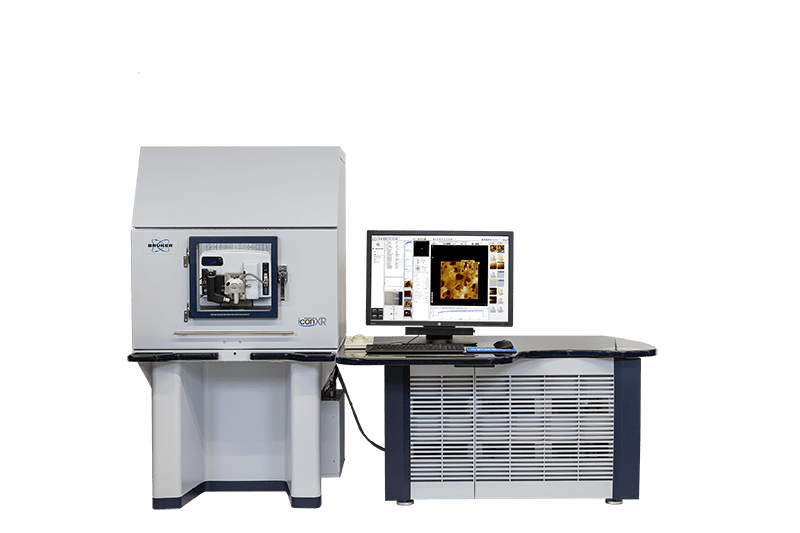
Atomic Force Microscopy (AFM)

Atomic Force Microscopy (AFM) measures surface topography of materials with sub-nm vertical resolution. The technique delivers fast data, with simple scans requiring only a few minutes to complete.

Strengths

  • Best height resolution among surface topography techniques
  • High lateral resolution with specialized cantilever tips
  • Rapid measurement: possible to capture images within 10 min
  • Alternative force probes (including: electric, magnetic, piezoelectric, etc) accommodate advanced analytical modes

Limitations

  • Limited field of view. Maximum scan size is 100 µm x 100 µm
  • Roughness must be less than 10 microns
  • Size, shape, and cleanliness of the tip may obscure the results

Learn More

Atomic Force Microscopy Services

Atomic Force Microscopy

Atomic Force Microscopy (AFM) measures surface topography of materials with sub-nm vertical resolution. The technique delivers fast data, with simple scans requiring only a few minutes to complete.

Electrostatic Force Microscopy

Atomic Force Microscopy (AFM) measures surface topography of materials with sub-nm vertical resolution. The technique delivers fast data, with simple scans requiring only a few minutes to complete.

Kelvin Probe Force Microscopy

Atomic Force Microscopy (AFM) measures surface topography of materials with sub-nm vertical resolution. The technique delivers fast data, with simple scans requiring only a few minutes to complete.

Magnetic Force Microscopy

Atomic Force Microscopy (AFM) measures surface topography of materials with sub-nm vertical resolution. The technique delivers fast data, with simple scans requiring only a few minutes to complete.

Sample Requirements

Example Outputs

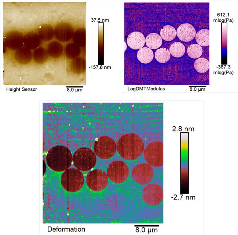
Quantitative Nanomechanical AFM images of carbon fibers encapsulated in epoxy: top left shows map of surface height; top right shows the log of the elastic modulus, with brighter areas on the carbon fibers corresponding to greater resilience and elasticity; bottom middle captures the deformation channel of this measurement, which shows the AFM tip compressing the fiber ends on the order of 2 nm.

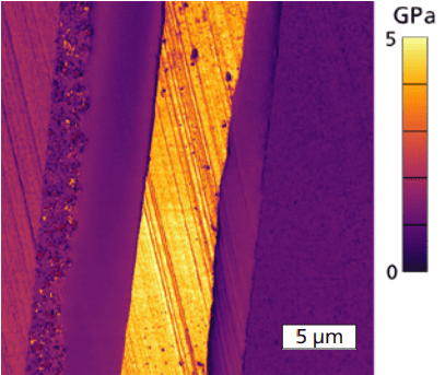
AFM map of the Elastic Modulus (GPa) measured across different films in a multilayer plastomer material using AM-FM mode.

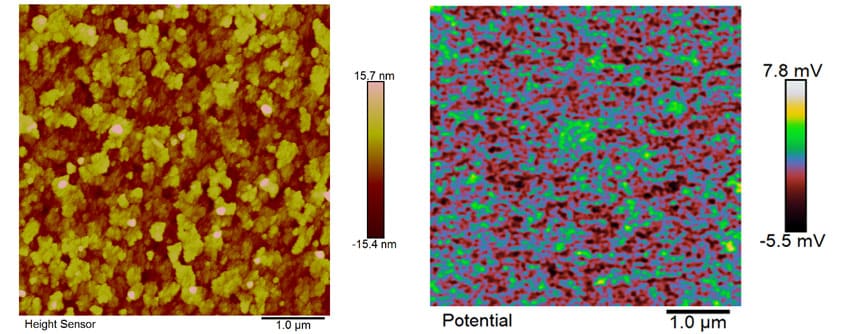
Tapping mode topography map on the left and Kelvin Probe Force Microscopy (KPFM) scan of surface potential at right. Scans were taken on Indium-doped Tin Oxide (ITO). KPFM can measure the work function of thin film surfaces and resolve minute differences in the surface potential using a specialized measurement mode which removes topographic contributions.

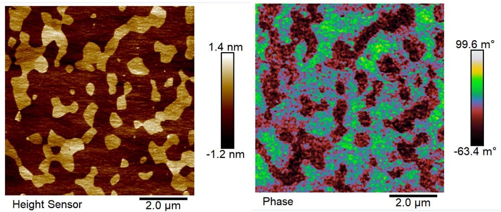
Topography and Electrostatic Force Microscopy (EFM) scan on titanium carbide alumina surface. Topography image on the left shows the higher TiC grains suspended in the Al2O3 matrix. On the right is an EFM phase image maps the difference in the probe tip’s electrical attraction to the TiC grains vs the alumina.

Instruments Used

Asylum Research Oxford Instruments Jupiter XR Atomic Force Microscope

Asylum Research Oxford Instruments Jupiter XR Atomic Force Microscope

The Jupiter XR from Oxford Instruments Asylum Research is the first and only large-sample AFM to offer both high-speed imaging and extended range in a single scanner. Jupiter provides complete 200 mm sample access and delivers higher resolution, faster results, a simpler user experience, and the versatility to excel in both academic research and industrial R&D laboratories.

  • Higher resolution than any other large-sample AFM
  • Extended range 100 um scanner is 5-20x faster than most other AFMs


View Instrument Brochure
Bruker  Nano Dimension (with Icon and FastScan Heads)

Bruker Nano Dimension (with Icon and FastScan Heads)

Whether using the Icon scanner with ultra-low noise and high accuracy, or employing the FastScan scanner for high scan rates, the Nano Dimension AFM from Bruker delivers exceptional ease of use and fast, high-resolution imaging and analysis.

View Instrument Brochure

How AFM Works

An AFM cantilever with a protruding ultra-sharp tip is raster scanned over the sample, which makes either intermittent or constant contact with the surface. The tip interacts with the sample, experiencing repulsive or attractive inter-atomic forces. A laser beam is reflected off the back of the cantilever onto a detector. As the cantilever scans, the detector monitors changes in the beam deflection. The z position of the cantilever shifts up or down to maintain a constant beam deflection and determine the vertical height of the surface.

Alternative advanced imaging modes allow for visualization and measurement of other material properties, such as: adhesion, modulus, charge distribution, work function, and magnetic domains (among others).