Focused Ion Beam Scanning Electron Microscopy (FIB-SEM)
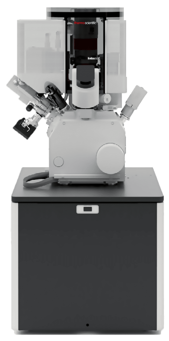
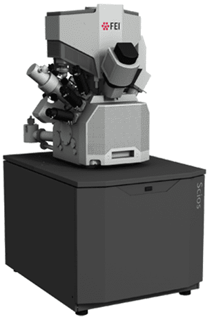
Like other high-resolution scanning electron microscopes, Focused-ion-beam scanning electron microscopes (FIB-SEMs) are used to produce 2D and 3D images of surface topography and can resolve nm-scale features on a sample surface.
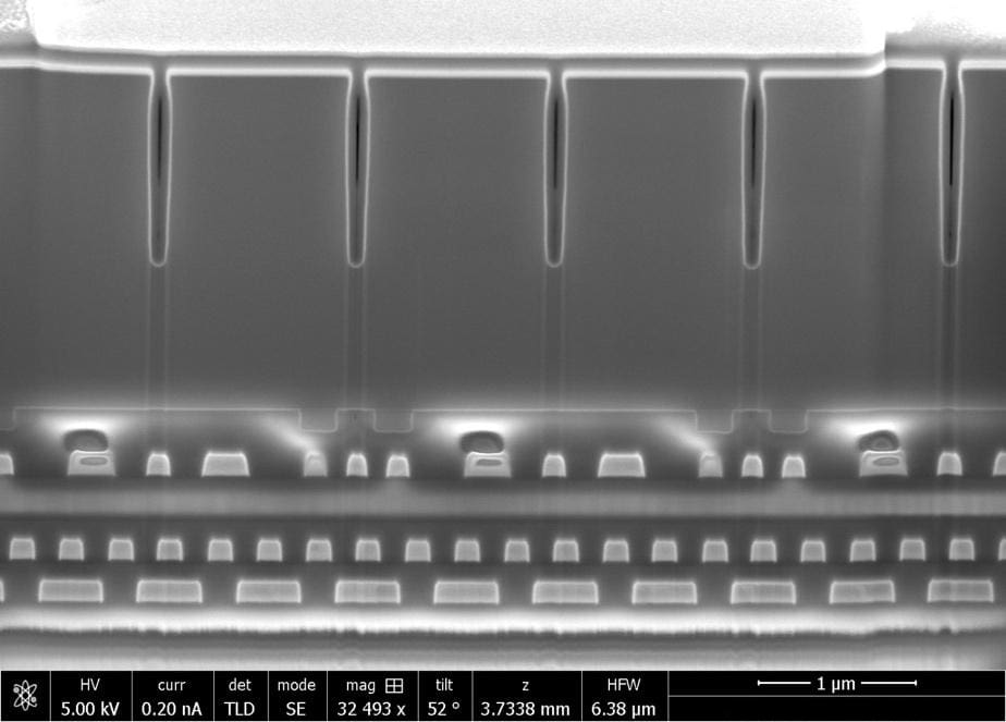
The FIB allows advanced analytical workflows such as cross-section, tomography, lithography, lamella prep, etc.
Strengths
- Ultra-high resolution imaging capabilities (limit resolution is < 1 nm)
- Able to image multi-modal, sub-surface, and 3D information
- FIB enables precise manipulation of sample in-situ: cutting, cleaving, trenching, exposing, and ion-welding of different fragments on the sample for imaging and analysis
Limitations
- Analysis is destructive
- Insulating materials impair precision of ion beam action and reduce imaging resolution


