Laser Scanning Confocal Microscopy (LSCM)

Laser scanning confocal microscopy (also called “VK” or “VKX” after the instrument used) is a nondestructive technique that generates 2D and 3D images of a sample surface. Covalent’s laser confocal microscopes can accomplish both broadband, white-light optical imaging and laser-confocal imaging.

Strengths

  • Controlled depth-of-field
  • 3D reconstructions and visualizations are possible with serial sectioning
  • Straightforward, relatively rapid data collection

Limitations

  • Optical properties of material determine end data quality
  • High-energy laser source can cause damage to live cells and tissues

Learn More

Laser Scanning Confocal Microscopy Services

Laser Scanning Confocal Microscopy

Laser scanning confocal microscopy (also called “VK” or “VKX” after the instrument used) is a nondestructive technique that generates 2D and 3D images of a sample surface. Covalent’s laser confocal microscopes can accomplish both broadband, white-light optical imaging and laser-confocal imaging.

Sample Requirements

Example Outputs

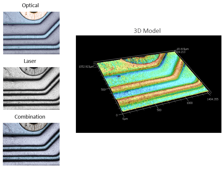
Images captured from the white light source (optical) and laser, as well as the combined image showing how the system captures highly accurate depth contrast as well as the true color of the different pieces of the sample. Pictured at left is a 3D model generated from the height profile of the sample surface; in this image, contrast and color is keyed to height instead of true sample color

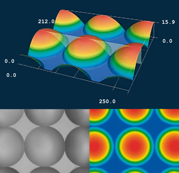
3D models generated from LSCM scans of microlenses, showing radius and circularity

From: Keyence

Instruments Used

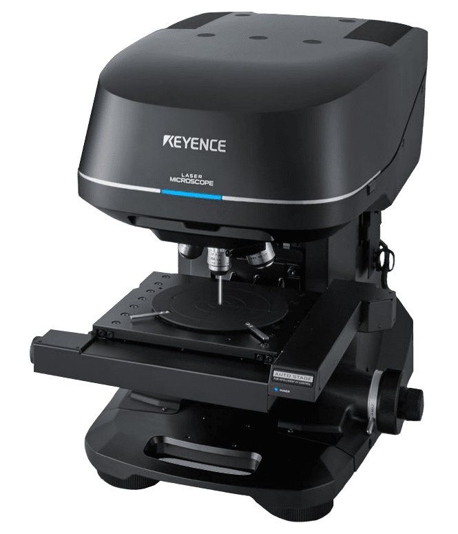
Keyence VKX-1100 Microscope

Keyence VKX-1100 Microscope

  • Lateral Scan Range: 100 mm x 100 mm
  • Vertical Stage Range: 70 mm
  • Lateral Resolution: < 1 µm
  • Vertical Resolution: < 1 µm (nm-scale possible in some cases)
  • Magnification Limit: up to 28,800X
View Instrument Brochure

How LSCM Works

For Laser-Confocal Microscopy, a laser source beam is passed through a set of optics which include narrow pinholes. The effect of these pinholes is to provide a very shallow depth of field such that only light which is reflected from near the exact focal plane of the final lens will reach the detector.

The microscope captures a series of vertical slices (in z-dimension) to build up a 3-dimensional profile at the illuminated beam spot. By scanning this spot in a raster-pattern laterally (in x-y plane), the system generates an image profile of the sample surface topography.

Nanometer-scale resolution can be achieved, depending on the focusing lens used for the measurement.