Nuclear Magnetic Resonance Spectroscopy (NMR)
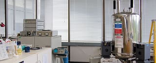
Nuclear Magnetic Resonance Spectroscopy Main Image
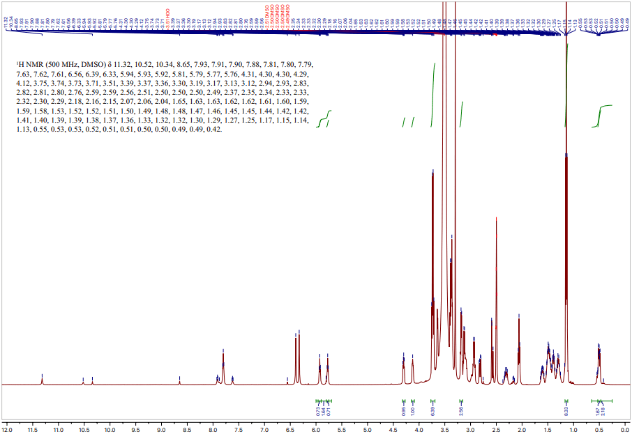
NMR spectrum generated from a 1H-NMR measurement.

Nuclear magnetic resonance spectroscopy (NMR) is a chemical analytical technique used to assay the composition and chemical structure of solutions, solids, mixtures, and macromolecules.

Due to its ability to capture dynamic molecular behavior, it can also be used to characterize reaction kinetics, real-time structural rearrangements, substrate binding and catalysis, and many other processes.

Strengths

  • Non-invasive and non-destructive (once isotopes are fully integrated)
  • Can be quantitative when used with reference / control samples
  • High precision measurement
  • Optimized for nonselective molecular analysis

Limitations

  • Sample preparation to use isotopic forms
  • Less sensitive to composition than mass spectrometry and other chemical analytical techniques
  • Limited to analyzing NMR-active nuclei

Learn More

Nuclear Magnetic Resonance Spectroscopy Services

Nuclear Magnetic Resonance Spectroscopy

Nuclear magnetic resonance spectroscopy (NMR) is a chemical analytical technique used to assay the composition and chemical structure of solutions, solids, mixtures, and macromolecules.

Due to its ability to capture dynamic molecular behavior, it can also be used to characterize reaction kinetics, real-time structural rearrangements, substrate binding and catalysis, and many other processes.

Sample Requirements

Example Outputs

NMR spectrum generated from a 1H-NMR measurement. Each peak represents a distinct electrochemical environment surrounding the NMR-active H nuclei; the chemical shifts (x-axis) associated with each peak can be used to identify functional groups present, listed in the top left. Taken together, these shifts then allow experts to characterize the molecular structure of the analyte.

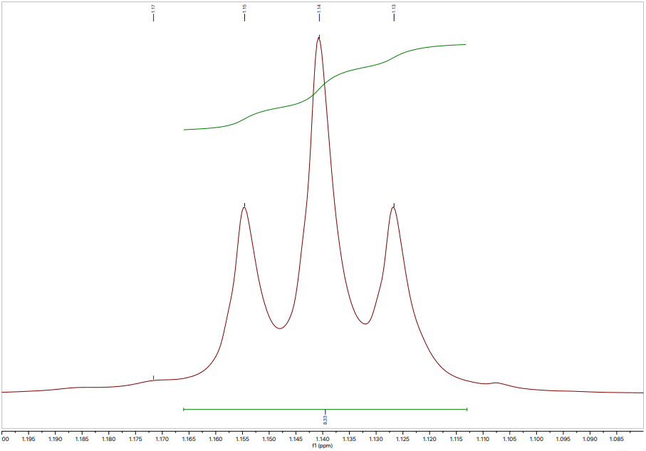
To ensure chemical shift is accurately measured and to allow precise correlative analysis for molecular structure determination, high resolution spectra are captured at each peak. Shown below is a multiplet peak isolated from the above total spectrum, with measured chemical shift and peak-splitting annotations that inform the chemical structure.

Instruments Used

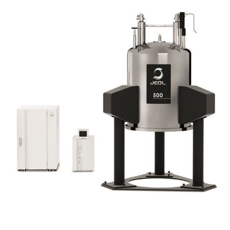
JEOL ECZ500R 500MHz NMR Spectrometer

JEOL ECZ500R 500MHz NMR Spectrometer

  • Magnetic Field Frequency: 500 MHz
  • Multi-Frequency Drive System: single and multiple nuclei experiments
  • Control Time Resolution: 5 ns (for phase, amplitude, and frequency modulation)
  • Wide Dynamic Range: up to 1 : 3,400,00
  • Sensitivity enhanced by 10% over past NMR systems
  • Probe Options:
    • ROYAL HFX Probe
    • SuperCOOL Probe- cools sample to LN2 temperature and improved sensitivity
    • 3.2 mm HX MAS Solid-state NMR Probe + Extended Variable Temperature Range
    • 3.2 mm HXY MAS Solid-state NMR Probe
    • Diffusion NMR probe
View Instrument Brochure
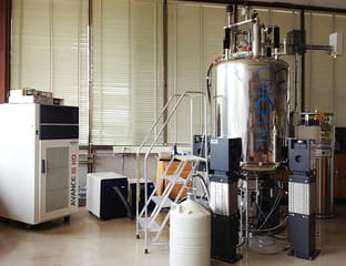
Bruker Avance III HD 600MHz NMR Spectrometer

Bruker Avance III HD 600MHz NMR Spectrometer

  • Magnetic Field Frequency Range: 5 to 650 MHz
  • Frequency Resolution: < 0.005 Hz
  • Phase Resolution: < 0.006°
  • Magnetic Field Strength: 14.1 Tesla
  • Probe Options:
    1. Multinuclear Prodigy CryoProbe – standard liquid samples
      – Temperature Range: -40 °C to +80 °C (for solution state samples only)
    2. Bruker SmartProbe – lower-frequency nuclei (as low as 109Ag), 1H/19F decoupling, and correlational (2D / 3D) NMR experiments
      – Temperature Ranger: -150 °C to +150 °C
  • High-power decoupling up to 15 kHz MAS (for solid-state NMR)
View Instrument Brochure
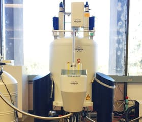
Bruker Avance III HD 500MHz NMR Spectrometer

Bruker Avance III HD 500MHz NMR Spectrometer

  • Magnetic Field Frequency Range: 5 to 500 MHz
  • Frequency Resolution: < 0.005 Hz
  • Phase Resolution: < 0.006°
  • Magnetic Field Strength: 11 Tesla
  • Probe: Multinuclear Prodigy CryoProbe and Bruker SmartProbe (same as above)
  • High-power decoupling up to 15 kHz MAS (for solid-state NMR)
Varian Inova 500 MHz Spectrometer

Varian Inova 500 MHz Spectrometer

  • 3 Radiofrequency channels with independent waveform generators
  • Optimized for 3D NMR experiments on 1H, 13C, 15N nuclei (‘HCN’) and dual-channel broadband experiments
  • Probe: 5mm Nalorac IDTG Triple-resonance Probe
  • Temperature Range: -100 °C to +150 °C

How NMR Works

NMR utilizes a strong, static magnetic field along with a weakly oscillating field surrounding the sample to induce and measure changes in the magnetic properties of select atomic nuclei.

Before the NMR measurement, the sample is treated and target atomic species are replaced with electromagnetically active isotopes. The isotopic atoms undergo spin-state polarization in the NMR as the weak field is turned on and off, and the energy and time associated with spin-state relaxation are measured. These values reflect the electromagnetic environment of each isotopically tagged atom;  which can be correlated to the overall molecular structure and composition.

Polarization is induced multiple times during a single NMR measurement, and differential field strengths permit 1D- and  2D-analytical modes, in which 1 or 2 atomic species are analyzed serially or simultaneously. This allows for quantitative molecular structure determination, and tracking of dynamic structural changes due to chemical processes.

Most NMR is done with liquid samples; however solid state NMR can be done with specialized equipment. There is also a variation of NMR done at low temperatures.