Photo-induced Force Microscopy (PiFM)

Photo-induced Force Microscopy Main Image

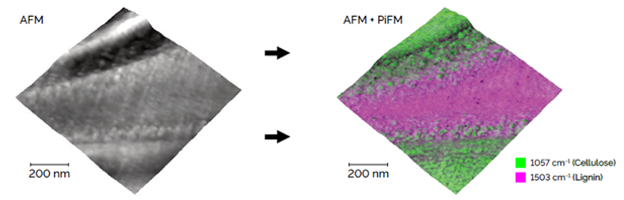
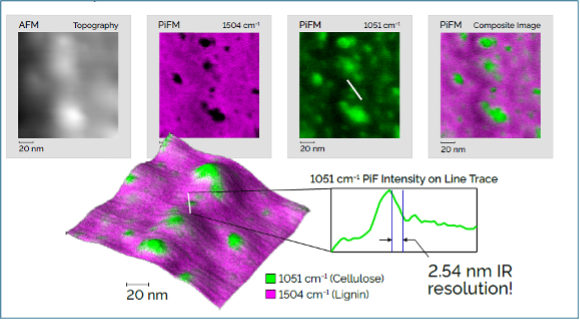
3D visualization of the AFM topography of a cell wall from an ultra-thin cross section of spruce wood. An overlay of two PiFM images the chemical composition of the surface where lignin and cellulose mix. This PiFM overlay reveals how the materials are distributed, and it shows how some of the topographic features are related to the local chemistry. Scan dimensions: 1 µm x 1 µm x 0.034 µm.

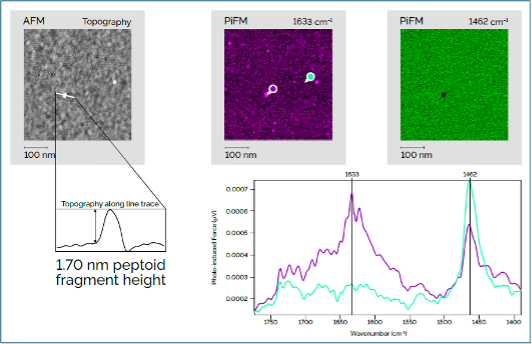
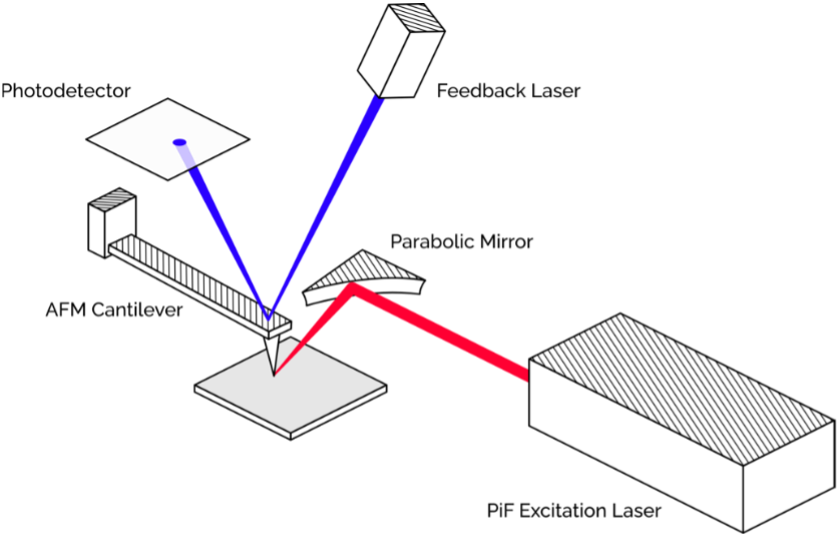
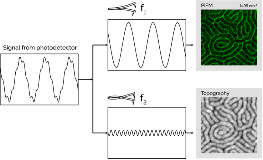
PiFM is a surface chemical analysis that utilizes tunable IR lasers combined with the AFM technique to provide nanoscale spatial resolution of topography and chemistry at ambient air conditions.

Strengths

  • Surface chemistry and topography at nanoscale resolution
  • Ability to measure chemical information (not elemental)
  • Non-contact and non-destructive, even more than SEM/EDS
  • Ability to measure in ambient air environments
  • Full-size wafer compatibility
  • Minimal or no sample preparations
  • Ease of use (vs. similar techniques) for nanoscale chemical measurements.

Limitations

  • Not well suited to pure metals and some 2D materials without IR-active peaks.
  • Accessibility of the sample surface is limited by the probe’s dimensions.
  • Due to the mechanical scanning, the imaging mode can be considered slow compared to optical and even electron microscopes.

Learn More

Sample Requirements

PiFM Example Outputs

PiFM measurement on a wood cell

This image presents typical results measured by PiFM:

  1. IR spectral data – this is exactly the kind of output for the chemical information that users familiar with FTIR would expect to see.
  2. AFM images & topography
  3. PiF-IR images & Chemical Mapping
    • Single WN (wavenumber) image
    • Multiple WN images overlaying

Figure1: Scan dimensions: 150x150x 10.5nm. A zoomed-in region of the spruce wood cell wall. PiFM images show the chemical distribution of lignin and cellulose on the surface. A line trace plotting the intensity of the data in the green image shows IR spatial resolution of less than 5nm. © Molecular Vista

Identification of organic contamination

In this example, identifying a peptoid with a height of 1.7 nm chemically and clearly distinguishing its spectra from those of its substrate will be extremely challenging. Likely, FIB-SEM and TEM are techniques capable of providing the necessary resolution and height measurement, but the EDS performed with TEM may not be able to chemically identify organic molecules like peptoids.

Figure 2: Scan dimensions: 500x500x1.7 nm. A PiFM image was taken at 1633cm-1, which should highlight any peptoid molecules present, like one shown on the scan of only 1.7nm tall. The black spot in the green PiFM image shows that the fragment is sitting on top of the substrate material. © Molecular Vista

PiFM Instruments Used

VISTA 150 by Molecular Vista

VISTA 150 by Molecular Vista

PiF Laser: QCL (770 – 1840, 1995 – 2395 cm−1)

Stage and scanner

Sample stage travel: 150 mm × 150 mm square.

Scan size: 90 µm × 90 µm.

Dual Z Feedback: 12 µm z-scanner (sample) with 600 nm fast-z scanner (tip) provides both high bandwidth and a large z-range

Functionality

Imaging modes: Non-contact AFM, PiFM, KPFM, cAFM, nano DMA, FvD (force vs distance) mapping.

Spectroscopy modes: PiF-IR, FvD.

There are two basic modes:

  • Surface Mode: measures up to 20 nm of depth of material
  • Bulk Mode: measures up to 1000 nm of depth of material

How PiFM Works

A pointed, metal-coated probe in PiFM – whose tip radius can be as small as 20 – 30 nm after the metal coating is applied – is set very close to the surface of the sample. The area immediately underneath the tip is illuminated by an exactly focused laser beam, which creates a localized electromagnetic near field. The near field then acts on the sample’s molecule, creating localized polarization based on the optical and chemical characteristics of the material.

Figure 3: Simple PiFM diagram. An excitation laser shown onto the sample surface allows photo-induced force detection via the AFM © Molecular Vista

This interaction between the tip and the polarization sample generates an extremely minute attractive dipolar force, which we refer to as the photo-induced force. The laser is modulated at a specific frequency so that the AFM probe can act as a mechanical amplifier of the photo-induced force. In scanning the tip across the surface and measuring these forces continuously, PiFM generates high-resolution maps that register surface topography and spatially resolved chemical contrast in parallel. We can keep the tip stationary and sweep the laser to collect a PiF-IR point spectrum.

Figure 4: The raw signal from the AFM photodiode has both the topography and PiFM signals combined. These two signals correspond to eigenmodes of the cantilever which allows independent detection after the signals are separated © Molecular Vista

As opposed to traditional optical microscopy, PiFM does not suffer from the diffraction limit of light and can have spatial resolutions of a few nanometers. This allows one to image at the molecular and nano scales, to differentiate between various species of chemicals, and to investigate heterogeneous materials at a degree of refinement impossible to reach with most other methods of spectroscopy.

Learn more about using Photo-induced Force Microscopy services today!