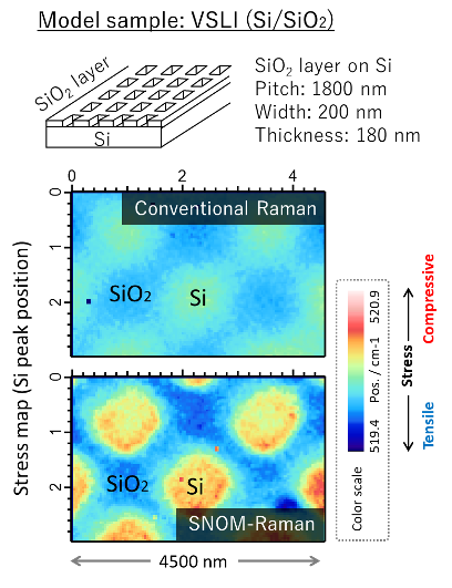
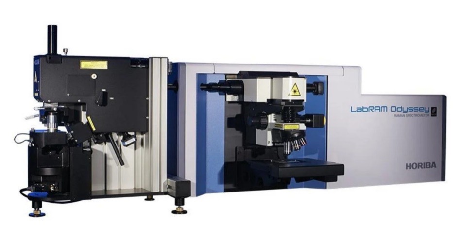
Scanning Near-field Optical Raman Microscope (SNOM-Raman)

Scanning Near-field Optical Raman Microscope Main Image

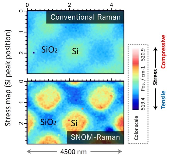
SNOM-Raman combines the capabilities of Scanning Near-field Optical Microscopy (SNOM) with Raman spectroscopy. It allows Raman spectroscopy to be performed with a much higher spatial resolution than traditional Raman microscopy.

Application areas: Semiconductors, Materials science

Strengths

  • Nanoscale spatial resolution, far beyond the diffraction limit of conventional Raman spectroscopy

Limitations

  • Maximum scan size is 0 µm x 3.0 µm
  • Roughness must be less than 1 µm

Learn More

Sample Requirements

Sample size: 10 mm × 10 mm, <10 mm thick

SNOM-Raman Example Outputs

The stress map on model Si device, in which grid silicon dioxide layer is deposited on a Si wafer. As seen here, the map of SNOM-Raman showed larger stress dynamic range owing to the increase of spatial resolution, and donuts-shaped compressive stress to exposed Si appears, indicating local strain concentration at Si-SiO2 interface.

SNOM-Raman Instruments Used

Horiba AFM-Raman (Horiba)

Horiba AFM-Raman (Horiba)

High spectral resolution

Ultimate spectral resolution performance, multiple gratings with automated switching, wide spectral range analysis for Raman and PL.

High spatial resolution

Nanoscale spectroscopic resolution (down to 10 nm) through Tip Enhanced Optical Spectroscopies (Raman and PhotoLuminescence).

Multi-technique / Multi-environment

Numerous SPM modes including AFM, conductive and electrical modes (cAFM, KPFM), STM, liquid cell and electrochemical environment, together with chemical mapping through TERS/TEPL. Full control of the 2 instruments through one workstation and a powerful software control, SPM and spectrometer can be operated simultaneously or independently

View instrument brochure

How SNOM-Raman Works

SNOM-Raman is a spectroscopic technique that uses near-field light generated from a subwavelength aperture. A focused laser interacts with the sample through a tiny aperture or tip, and the scattered light is analyzed to produce high-resolution Raman spectra.

Possible measurements:

Chemical composition
Stress distribution

Technical specifications:

Spatial resolution: 100 nm

Learn more about using Scanning Near-field Optical Raman Microscope services today!